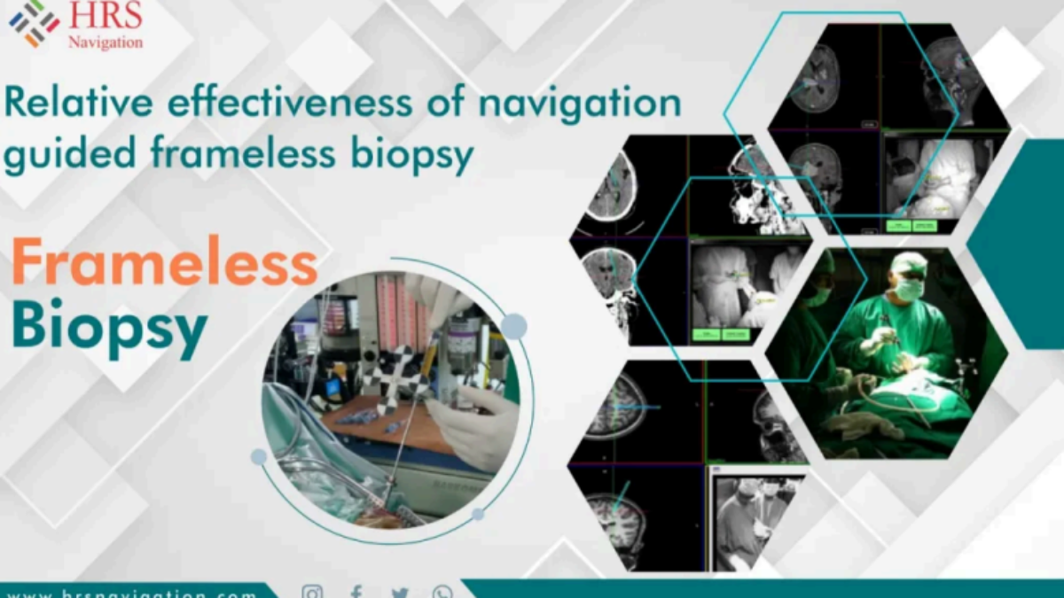
Over the past four decades, brain biopsy procedures have continuously evolved in line with advances in brain imaging. Among these, frame-based stereotactic interventions have long been considered the gold standard for obtaining brain biopsy specimens.
What is Frame-Based Stereotactic Biopsy?
- A rigid head frame is affixed to the patient’s skull before the imaging.
- The frame remains an integral part of the biopsy, ensuring high precision, accuracy, and diagnostic yield.
However, despite its reliability, this method comes with several limitations.
Limitations of Frame-Based Stereotactic Biopsy
Here are the key challenges associated with the frame-based technique:
- 🧷 Discomfort due to bulky head frame and fixation pins
- ⏳ Longer pre-operative time due to frame placement, imaging, and coordinate calculation
- 🧮 Complex calculations to determine stereotactic entry points
- 🚧 Obstructed surgical field because of the frame’s physical structure
- 🧫 Increased infection risk at pin fixation points
- ❌ No real-time needle visualization or feedback during biopsy
- ⏱️ Prolonged surgical time
The Evolution: Enter Frameless Stereotactic Biopsy
Thanks to continuous advances in image-guided neurosurgery, a more flexible and equally accurate approach has emerged—Frameless Stereotactic Biopsy.
Why Frameless is the Preferred Modern Alternative
- ✅ No frame fixation needed—eliminates discomfort and pin site infections
- ✂️ Smaller incisions—less invasive and quicker healing
- 🧠 Real-time imaging of the biopsy needle inside the brain
- 📍 Dynamic trajectory planning—from entry to lesion target
- 🧭 Navigation via software—enables fusion of multiple radiographic images
- 🧾 Registration based on anatomical landmarks—eliminates need for external fixation
- 🩺 Simultaneous monitoring of needle depth and location for assured precision
How It Works: Simplified Surgical Navigation
The frameless procedure uses:
- Pre-operative scans for landmark registration
- Custom trackers and navigation software for image fusion
- Dynamic trajectory planning for a targeted approach
- Real-time feedback on biopsy needle position and depth
This system allows neurosurgeons to perform the biopsy with greater confidence, reduced invasiveness, and shorter operative times—while maintaining diagnostic accuracy equivalent to frame-based methods.
Conclusion
Frameless stereotactic biopsy is rapidly becoming a standard technique in modern neurosurgery. By overcoming the limitations of traditional frame-based procedures, it delivers:
- Comfort for the patient
- Operational ease for the surgeon
- Diagnostic precision backed by real-time imaging
In a field where precision is critical, frameless stereotactic biopsy stands out as a reliable, safe, and effective solution.
